Age-Related Macular Degeneration (AMD)
Macular Degeneration (AMD) is the leading cause of irreversible vision loss in people over 60, affecting nearly 20 million Americans. The risk of developing AMD increases with age, and by age 75, the likelihood rises to 1 in 3. There is also a juvenile form of macular degeneration called Stargardt disease, which affects younger individuals and has different causes and treatments.
While there are treatment options to maintain sight and slow disease progression, AMD is currently an incurable condition. AMD leads to the deterioration of the macula, the central part of the retina responsible for detailed vision, such as reading, recognizing faces, and perceiving color and contrast. As the macula deteriorates, vision may become blurry, wavy, or eventually lost entirely in the center of the visual field, while peripheral vision remains unaffected.
Stages of Macular Degeneration
Regular eye exams are crucial for early detection and managing AMD progression.
- Sub-clinical AMD: Early stage where dark adaptation is impaired, but no visible changes to the macula.
- Early AMD: No noticeable vision loss, but medium-sized drusen (yellow deposits) may appear.
- Intermediate AMD: Some vision loss may occur, with larger drusen or pigment changes.
- Late AMD: Significant vision loss due to advanced dry AMD (geographic atrophy) or wet AMD.
![]()

Types of Macular Degeneration
- Dry AMD: The most common form, accounting for 85-90% of cases.
- Geographic Atrophy (GA): The advanced stage of dry AMD.
- Wet AMD: Less common, affecting 10-15% of cases, but often more severe.
- Stargardt Disease: A form of macular degeneration affecting younger people.
Dry Age-Related Macular Degeneration
Dry is the most common form of age-related macular degeneration, affecting about 80-90% of those with the condition. It occurs when the central part of the retina, called the macula, thins and tiny deposits called drusen accumulate underneath it. This leads to central vision loss, with the severity depending on the location and amount of retinal thinning.
Early stages of dry AMD may cause minimal vision problems, but as the condition progresses, it can cause blurry central vision, difficulty reading, and trouble seeing in low light. The central vision may also develop blank spots, and straight lines might appear wavy. Peripheral vision is typically unaffected.
Dry AMD tends to progress more slowly than the "wet" form but can eventually turn into wet AMD over time.
Photobiomodulation Treatment
LumiThera offers the first and only FDA-approved treatment for early to intermediate dry age-related macular degeneration (AMD). This non-invasive, red-light therapy, called photobiomodulation (PBM), is delivered using the Valeda® Light Delivery System to slow the progression of dry AMD, improve vision, and enhance quality of life.
PBM works by stimulating the mitochondria in the retina to boost metabolic function, reduce inflammation, and protect against further vision loss. In clinical trials, PBM treatment resulted in significant improvements in vision and a reduction in the progression of dry AMD, including a lower risk of developing severe forms like wet AMD or geographic atrophy, which leads to permanent vision loss.
The treatment consists of a series of 9 sessions per eye over 3-5 weeks, with follow-up treatments every 4 months. Each session lasts about 30 minutes, with the therapy taking less than 5 minutes per eye. It is non-invasive, painless, and requires no special preparation, making it a convenient option for patients looking to preserve their vision.
Wet Age-Related Macular Degeneration
Wet macular degeneration (nAMD) is a more advanced form of age-related macular degeneration (AMD), affecting 10-15% of AMD patients. It is characterized by the growth of abnormal blood vessels beneath the retina and macula. These new blood vessels, known as choroidal neovascularization (CNV), can leak blood and fluid, causing the macula to bulge and potentially leading to rapid vision loss and distortion.
Symptoms
Patients with wet AMD may notice dark spots in their central vision or experience straight lines appearing wavy. Central vision loss can occur quickly, but side vision typically remains unaffected. In some cases, patients may not notice symptoms until the condition has progressed. Regular eye exams are crucial, especially for those at high risk, as early intervention can help prevent further vision loss.
Risk Factors
If CNV develops in one eye, the risk of the condition developing in the other eye is high. Factors that increase the risk of CNV include the presence of large drusen, pigmentary changes in the retina, and high blood pressure. A combination of these factors significantly raises the risk of CNV in the second eye.
Treatment Options
Unlike dry AMD, which can often be managed with lifestyle changes, supplements, and photobiomodulation therapy, wet AMD requires early and aggressive treatment. The main goal of treatment is to control the bleeding and prevent further retinal damage.
The most common treatment for wet AMD involves anti-VEGF (vascular endothelial growth factor) drugs, which target the abnormal blood vessels that cause bleeding. These drugs stop the growth of new blood vessels and reduce leakage. Common FDA-approved anti-VEGF medications include:
- Bevacizumab (Avastin)
- Ranibizumab (Lucentis)
- Aflibercept (Eylea)
- Brolucizumab (Beovu)
- Faricimab (Vabysmo)
These medications are injected into the eye at regular intervals. The frequency of injections depends on the patient's condition and the specific drug used, with many patients requiring injections every 4-6 weeks to maintain effectiveness. While anti-VEGF drugs do not restore vision, they can help prevent further vision loss.
Injections Risks
Though effective, anti-VEGF injections carry some risks, including ocular inflammation, increased eye pressure, endophthalmitis (serious infection), retinal detachment, conjunctival hemorrhage. It's important to discuss these risks with your doctor.
Geographic Atrophy
Geographic Atrophy (GA) is an advanced stage of dry age-related macular degeneration (AMD) that causes permanent vision loss. It occurs when cells in the retina die, creating distinct areas of damage, known as lesions. These lesions can blur central vision, making everyday tasks like reading, driving, or seeing in low light more difficult. Over time, these lesions may expand and affect the fovea, the center of the retina responsible for sharp vision, leading to blind spots and loss of central vision.
Risk Factors
- About 30% of people with dry AMD will develop GA.
- The risk increases as dry AMD progresses from early to intermediate stages.
- Factors such as smoking, poor visual acuity, and chronic health conditions like obesity, cardiovascular disease, diabetes, and high cholesterol can increase the risk of GA.
Wet AMD and Geographic Atrophy
- Up to 37% of people with wet AMD may develop GA within two years, and by 7.3 years, nearly all wet AMD patients may also develop GA.
- GA can develop during the later stages of dry AMD or after wet AMD, even after treatment with anti-VEGF injections. It’s important to manage AMD and related health conditions to reduce the risk of GA progression.
Treatment Options
For more than a decade there was no successful way to treat geographic atrophy (GA), until in 2023, when the Food and Drug Administration (FDA) approved two intravitreal injection treatments:
- Pegcetacoplan (Syfovre)
- Avacincaptad pegol (Izervay)
Syfovre and Izervay are designed to inhibit and provide comprehensive control of the complement cascade, part of the body’s immune system. By regulating that overactivated part of the immune system in your eye, injections may help slow the progression of GA.
AMD Fact Sheets
Download the following documents to learn more about age-related macular degeneration.
PDF Fact Sheet (1)
PDF Fact Sheet (2)
Large-Print Version
Spanish Translation
Risk Factors
You are more likely to develop AMD if you:
- eat a diet high in saturated fat
- are overweight
- smoke cigarettes
- are over 50 years old
- have hypertension, high cholesterol or heart disease
- have a family history of AMD
Testing & Diagnosis
To assess your vision and the health of your eyes, several tests may be performed to detect AMD in addition to measuring visual acuity.
- Amsler Grid Test: This test checks your central visual field to detect any distortions or blind spots, which are common signs of macular degeneration.
- Dilated Eye Exam: Your doctor will apply special eye drops to dilate your pupils, allowing them to closely examine your retina using specialized lenses.
- Fluorescein Angiography (FA): In this test, a yellow dye (fluorescein) is injected into a vein, typically in your arm. A special camera takes detailed photos of your retina as the dye moves through the blood vessels, helping to identify abnormal blood vessel growth beneath the retina.
- Optical Coherence Tomography (OCT): This non-invasive test uses a scanning laser to capture high-resolution images of your retina, offering a detailed view of the macula and helping to monitor changes or damage.
These tests are essential in diagnosing macular degeneration and monitoring disease progression, allowing your doctor to recommend the best treatment options.
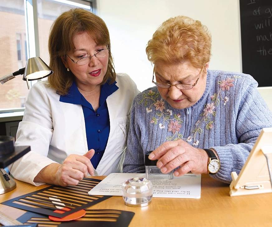
Low Vision Rehabilitation
Schedule an Age-Related Macular Degeneration Consultation with Colorado Retina Associates
As the premier retina practice of the Rocky Mountains, Colorado Retina Associates provides advanced diagnostic care and treatment for Age-Related Macular Degeneration. Schedule a consultation today with one of our retina specialists in the Denver, Boulder, Aurora, Littleton, and Lakewood areas.